Fundamentals of Psychopharmacology

Fundamentals of Psychopharmacology

Understanding psychopharmacology is foundational to providing mental healthcare. Review pathways, brain regions, receptors, and functions associated with various neurotransmitters that are thought to play a role in psychiatry.

Introduction to Psychopharmacology

Psychopharmacology explores how neurotransmitters in the nervous system affect physiological processes. Neurotransmitters can have diverse effects on physiological processes including mood, behavior, cognition, and movement. Abnormal levels of neurotransmitters are thought to play a role in various diseases, including major depressive disorder (MDD), bipolar I disorder, and schizophrenia. The study of psychopharmacology aims to understand neurotransmitter imbalances and the potential effects therapeutic agents may have.1,2

Visualizing the synthesis, localization, and metabolism of certain neurotransmitters may help to understand how various symptoms can manifest in psychiatric disorders. Healthcare providers can learn how these neurotransmitters are thought to be modulated to better understand the potential impact they may have in psychiatry.1

*Please note that this article provides only a theoretical overview, and the clinical relevance of pre-clinical studies is unknown. 

Neurotransmitters

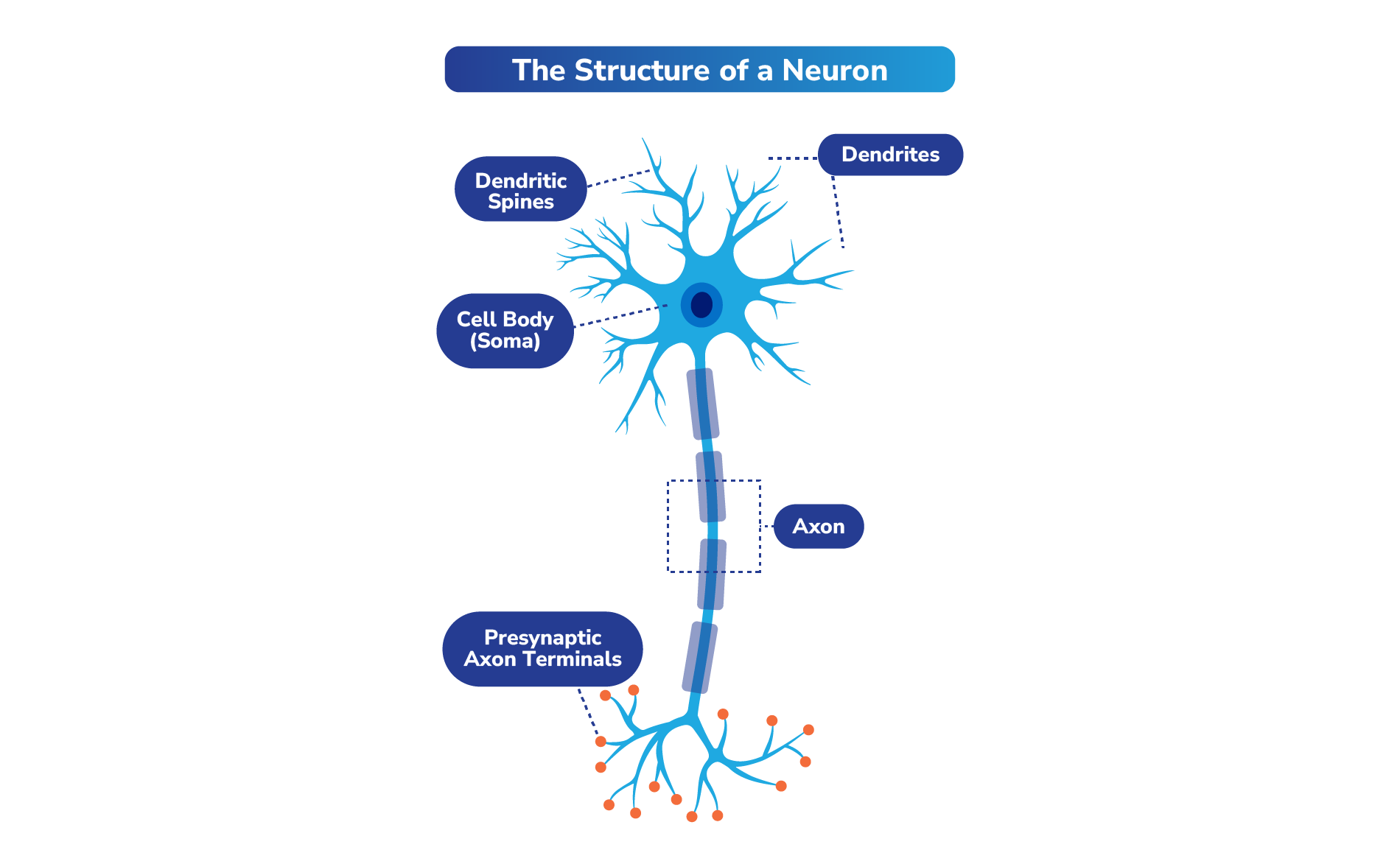
Neurotransmitters are chemical signaling molecules in the nervous system that are transmitted through neurons (cells in the nervous system, or nerve cells). It has been estimated that an average of 86 billion neurons make up the brain, forming a network of connections responsible for our thoughts, emotions, and actions.3

Structure of a neuron

    While the structures of neurons can vary depending on their role and location in the brain, nearly all neurons have 3 essential parts: a cell body (soma), an axon, and dendrites.1 The unique structure of neurons allows them to receive, process, and transmit information to one another. The synapse is the point of contact between the axonal projection of the neuron and other cells through which neurotransmitters travel.4

    How Neurotransmitters Work

    Neurotransmitters are key information carriers throughout the central and peripheral nervous system. They are endogenous chemical messengers that signal neurons and other cell types, affecting functions such as emotions, thoughts, memories, movement, and sleep.2

    Neurotransmission is the process by which information is shared between neurons through neurotransmitters. A neuron sending a signal (ie, presynaptic neuron) releases neurotransmitters into the synapse. These neurotransmitters bind to receptors on the surface of a receiving neuron (ie, postsynaptic neuron) and can either activate or block cellular functions downstream. The mechanisms underlying neurotransmission involve both chemical and electrical signals. Within a neuron, electrical impulses, or action potentials, are sent from one part of the cell to another via the axon.1

    Once an action potential is passed down to the axon terminal, the electrical signal is converted to a chemical one as neurotransmitters are released into the synapse to signal adjacent neurons.1,4

    Chemical information is converted by these receptors and associated proteins into electrical information by processes involving the activation of ion channels.5

    Neurotransmitters can be excitatory or inhibitory depending on whether they cause a depolarization or hyperpolarization of the neuronal membrane.6

    Types of Neurotransmitters

    Neurotransmitters can be classified into groups based on their chemical nature.2 Some common ones are listed below. 

    Monoamine neurotransmitters are thought to play a role in motor functions, emotional responses, motivation, and behavioral functions. Some monoamines include dopamine, serotonin, and norepinephrine.2

    • Dopamine is thought to participate directly or indirectly in most physiological functions, including motor control and cognition, occurring within the central nervous system (CNS).2
    • Serotonin is also thought to be involved in many physiological processes, including modulating sleep and wake states, gastrointestinal secretion and peristalsis, respiration, vasoconstriction, behaviors (feeding, aggression, mood/depression), and other neurological functions.2
    • Norepinephrine and epinephrine have roles as both hormones and neurotransmitters. As neurotransmitters, they are involved in the autonomic nervous system (ie, the “fight or flight” response). Norepinephrine is thought to play a role in alertness, sensory signal detection, regulation of emotions, memory, learning, and attention. Epinephrine is thought to have an impact in the “fight or flight” response by increasing heart rate, vasodilation, pupil dilation, and blood sugar levels.2

    Amino acid neurotransmitters include α-amino acids such as glutamate and γ-amino acids such as γ-aminobutyric acid (GABA). These neurotransmitters are thought to be involved with fundamental brain processes, including arousal, sleep, and consciousness.2,7

    • Glutamate is known as the predominant excitatory neurotransmitter in the CNS. It is the precursor to GABA. Glutamate is important for long-term potentiation, or long-lasting strengthening of synaptic connection,8 and may contribute to cognitive functions, including learning and memory formation.2
    • GABA is the main inhibitory neurotransmitter and can be formed by the conversion of glutamate to GABA in interneurons.2 GABA is thought to play a role in many functions, including behavior, motor control, mood, and sleep.9

    Other neurotransmitters: Acetylcholine was the first neurotransmitter identified in the peripheral nervous system and is responsible for muscle contraction within the neuromuscular system. In the CNS, acetylcholine is believed to play a role in consciousness, attention, learning, memory, sleep, and voluntary movement control.2

    Select Neurotransmitter Pathways and Receptors That May Play a Role in Schizophrenia, Bipolar I Disorder, and/or MDD

    Dopamine

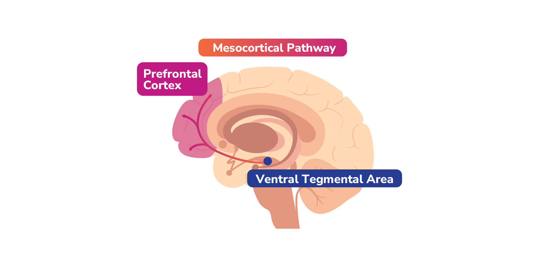
    Dopamine is a neurotransmitter produced in the dopaminergic neurons located in the substantia nigra and ventral tegmental area (VTA).10 In preclinical studies, it has been shown to be involved in the regulation of a range of physiological functions of the CNS, including motor control, feeding, affect, reward, sleep, memory, learning, and cognition.2,11

    Dopamine Pathways

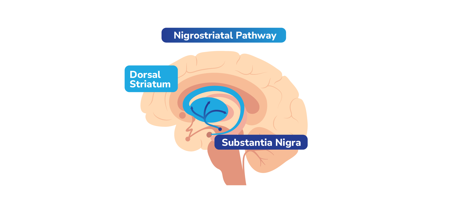
    Dopamine travels to different parts of the brain through 4 key pathways, each with different functions.1

    Mesolimbic pathway: Projects dopaminergic neurons from the VTA to the nucleus accumbens (NAc) in the ventral striatum.1

    Mesolimbic pathway

    This pathway is thought to be associated with regulating motivation, pleasure, and reward. Hyperactivity within this pathway is thought to be related to positive symptoms in schizophrenia, such as hallucinations and delusions. It also may be implicated in aggressive and hostile behaviors, particularly in schizophrenia and related disorders. On the other hand, a deficiency in the functioning of this pathway could potentially play a role in depressive symptoms, such as anhedonia.1

    Mesocortical pathway: The mesocortical pathway originates in the VTA and projects to the prefrontal cortex (PFC).1

    Mesocortical pathway

    This pathway is thought to regulate cognition and executive functions in the dorsolateral PFC, as well as emotions and affect in the ventromedial PFC. It is hypothesized that a deficit of dopamine activity within the mesocortical projections to both the dorsolateral and ventromedial PFC may be related to the cognitive, affective, and other negative symptoms of schizophrenia.1

    Nigrostriatal pathway: The nigrostriatal pathway originates in the substantia nigra in the brainstem and projects to the basal ganglia or striatum.1

    Nigrostriatal pathway

    A component of the extrapyramidal nervous system, the nigrostriatal dopamine pathway is believed to be responsible for controlling certain motor movements. A deficiency of dopamine in this pathway is thought to be involved in the development of movement disorders such as akathisia. On the other hand, hyperactivity of dopamine in the nigrostriatal pathway may be associated with hyperkinetic movement disorders such as chorea, dyskinesias, and tics.1

    Tuberoinfundibular pathway: The dopamine neurons in the tuberoinfundibular pathway project from the hypothalamus to the anterior pituitary.1

    Tuberoinfundibular pathway

    Dopamine neurons in the tuberoinfundibular pathway may play a role in the regulation of prolactin. When dopamine neurons in this pathway are active, prolactin release is inhibited. If these neurons are disrupted or affected by medications, prolactin levels may rise. Elevated prolactin levels may lead to breast secretions, loss of ovulation and menstrual periods, and potentially sexual dysfunction.1

    Dopamine Receptor Types

    Dopamine’s physiological effects are mediated by its interactions with dopamine receptors located across the brain.12 There are 5 known types of dopamine receptors categorized into 2 subfamilies: the D1-like and D2-like receptors.

    • D1-like receptors have an excitatory effect on the postsynaptic neuron. D1-like receptors include D1 and D5 receptors.13
    • D2-like receptors have an inhibitory effect on the postsynaptic neuron. D2-like receptors include D2, D3, and D4 receptors.13
    D2-like receptors are expressed both presynaptically and postsynaptically, while D1-like receptors are found to be expressed only postsynaptically.14 The D2 receptor is present in all 4 dopaminergic pathways.1 The basal nuclei (ie, basal ganglia), in particular, are rich in D2 receptors.15 Blocking D2 receptors in the mesolimbic pathway may lead to an inhibition of dopamine release that can potentially help reduce the positive symptoms of psychosis. D2 antagonism in the nigrostriatal and tuberoinfundibular pathways, however, is thought to be associated with extrapyramidal symptoms (EPS) and hyperprolactinemia.1

    The D3 receptor is mainly expressed in the limbic region of the brain with the highest concentrations found in the NAc, VTA, and the substantia nigra.13 In preclinical studies, it has been shown to be associated with regulating emotions, reward, motivation, and cognition.16 A pre-clinical study showed that the D3 receptor may also be associated with memory and attention.17

    Serotonin

    Serotonin is involved in many physiological processes. Dysregulation of serotonin is hypothesized to play a role in the pathology surrounding MDD and psychosis.18,19

    Serotonin Pathway

    In the CNS, serotonin, also known as 5-HT, is primarily produced in the raphe nuclei located in the brainstem. These cells have widespread connections throughout the brain and spinal cord.13

    The raphe nuclei include13:

    • The rostral nuclei, which project to most parts of the brain, including the cerebellum.
    • The caudal nuclei, which connect to the cerebellum, brainstem, and spinal cord.
    Together, these 2 clusters of serotonergic neurons innervate most of the CNS.20 

    Serotonin Receptor Types (Examples)

    There are at least 15 currently known subtypes of serotonin receptors, which are classified into 7 serotonin receptor classes, or subfamilies, based on their structure and pharmacologic action.13,21,22 Except for the 5-HT3 receptor subtype, which are ligand-gated ion channels, all serotonin receptor subtypes are G-protein-coupled receptors. The serotonin receptors that are thought to play a role in neuropsychiatric disorders include 5-HT1A, 5-HT2A, 5-HT2B, and 5-HT2C.13

    Example Serotonin Receptors and Their Functions

    Receptor Hypothesized Functions 
    5-HT1A receptor Highly expressed in the midbrain, limbic, and cortical regions. The 5-HT1A receptor may be involved in regulating mood and stress responses.23 Decreased 5-HT1A receptor binding may be implicated in depression.13,24
    5-HT2A receptor  

    Found to be widely distributed in the brain with relatively high expression throughout the cortex and can be found in the hippocampus, basal ganglia, and forebrain.1,23

    The 5-HT2A receptor may be associated with modulating cognition, perception, and mood.25,26 Some pre-clinical in vivo studies showed decreased 5-HT2A receptor binding in the cortex in patients with schizophrenia and patients with MDD.13,26,27 

    5-HT2B receptor  Found to be distributed at lower levels in the brain and peripheral tissues, the 5-HT2B receptor is thought to be involved with anxiety, appetite, and sleep.13 Clinical studies have shown that the 5-HT2B receptor may also play a role in all symptom domains of schizophrenia, including attention, social interactions, and learning and memory, as well as depressive behaviors.28,29
    5-HT2C receptor  Located in the choroid plexus, substantia nigra, and basal ganglia.13 The 5-HT2C receptor may be involved in regulating appetite, mood, and reward pathways.30 Dysregulation of the 5-HT2C receptor is thought to play a role in depression.13,22 

    Norepinephrine

    Norepinephrine, or “noradrenaline,” has a dual role as a neurotransmitter and a hormone. It is believed to be involved in alertness, sensory signal detection, and regulation of emotions, memory, learning, and attention.2 Norepinephrine is produced from the amino acid tyrosine through a series of enzymatic steps.1

    Norepinephrine Pathways

    In the central nervous system, norepinephrine has both ascending and descending projections. Ascending projections mostly originate in the locus coeruleus of the brainstem and extend to multiple regions of the brain. Ascending projections may help to regulate mood, arousal, and cognition. Descending projections extend down the spinal cord and may help regulate the pathways associated with pain.1

    Norepinephrine Receptors

    Norepinephrine is the primary neurotransmitter of noradrenergic neurons. Receptors that regulate norepinephrine neurotransmission include α1, α2A, α2B, α2C, β1, β2, and β3 receptors. All can be postsynaptic, while α2 receptors can also act as presynaptic autoreceptors.1

    Presynaptic α2 receptors regulate norepinephrine release through a negative-feedback regulatory signal. The binding of norepinephrine to these autoreceptors causes the neuron to turn off further release of norepinephrine. Certain medications may mimic the stimulation of this autoreceptor, thus preventing the release of norepinephrine. Other medications working as antagonists can have the opposite effect, increasing the release of norepinephrine.1

    Norepinephrine projections

    A = amygdala, BF = basal forebrain, C = cerebellum, H = hippocampus, Hy = hypothalamus, NA = nucleus accumbens, NT = brainstem neurotransmitter centers, PFC = prefrontal cortex, S = striatum, SC = spinal cord, T = thalamus.

    Monoamine Reuptake Mechanisms

    Certain enzymes and receptors play an important role in regulating free monoamine transmitter levels. Reuptake mechanisms via presynaptic transporters (ie, dopamine transporter receptor [DAT], serotonin transporter receptor [SERT], and norepinephrine transporter [NET]) allow for excess dopamine, serotonin, and norepinephrine to be transported back into their respective presynaptic neurons. Thereafter, each monoamine transmitter utilizes the vesicular monoamine transporter (VMAT2) receptor to be stored into the synaptic vesicles of their respective neurons for future use. The neurotransmitters that do not become packaged in synaptic vesicles are degraded in the neuron by the enzymes monoamine oxidase (MAO)-A or MAO-B or outside the neuron by catechol-O-methyl-transferase (COMT).1

    Monoamine reuptake mechanisms

    Adapted from Stahl SM. Stahl’s Essential Psychopharmacology. 4th ed. Cambridge University Press; 2013:89-96. COMT = catechol-O-methyl-transferase, MAO A/B = monoamine oxidase A/B, NT = neurotransmitter.

    Glutamate

    Glutamate is known as the main excitatory neurotransmitter in the CNS, with the ability to excite or “turn on” almost any neuron.1

    Glutamate Pathways 

    Glutamate pathways

    (a) Cortico-brainstem glutamate pathway; (b) Cortico-striatal/cortico-accumbens glutamate pathway; (c) Hippocampal accumbens glutamate pathway; (d) Thalamo-cortical glutamate pathway; (e) Cortico-thalamic glutamate pathway; (f) Direct cortico-cortico glutamatergic pathway; (g) Indirect cortico-cortico glutamatergic pathway 

    There are 6 glutamatergic pathways of particular interest in the field of psychopharmacology.

    Glutamate Pathways and Their Possible Downstream Impact


    Pathway DescriptionDownstream Impact
    Cortico-brainstem glutamate pathwaysProjecting from the cortical pyramidal neurons to brainstem neurotransmitter centers, these pathways include the raphe nuclei for serotonin, VTA and substantia nigra for dopamine, and the locus coeruleus for norepinephrine.This pathway plays a role in the release of monoamine neurotransmitters (norepinephrine, serotonin, dopamine). Direct innervation of monoamine neurons by excitatory glutamate neurons stimulates monoamine release within the brainstem. Opposingly, indirect innervation of monoamine neurons by excitatory glutamate neurons via GABA interneurons blocks monoamine release.1,31 These glutamate pathways may play a role in regulating dopamine release within the mesolimbic and mesocortical dopamine projections through a complex set of interactions. Interactions that contribute to dopamine hyperactivity are thought to play a role in the positive symptoms of schizophrenia.1
    Cortico-striatal/cortico-accumbens glutamate pathwaysThe pathways extend from the cortical pyramidal neurons to the striatal complex. They can project to the dorsal striatum (cortico-striatal pathway) or to the nucleus accumbens (cortico-accumbens pathway).These glutamate pathways travel down to the striatal complex and end on specific neurons that release GABA. The GABA neurons then signal the globus pallidus, another part of the striatal complex.1 These pathways may be involved in cognitive, motor, and affective functions, including motor control, sequence learning, and habit formation.32,33 
    Hippocampal-accumbens glutamate pathwayThis pathway projects from the hippocampus to the nucleus accumbens.Similar to the cortico-striatal and cortico-accumbens glutamate pathways, this pathway sends GABA neurons to a relay station located in the globus pallidus. Excessive glutamate output by glutamate neurons in this pathway that project to the mesolimbic dopamine neurons can also lead to dopamine hyperactivity, which may manifest as the positive symptoms of schizophrenia.1
    Thalamo-cortical glutamate pathwayThis pathway ascends from the thalamus and innervates pyramidal neurons in the cortex.It helps to communicate information between the cortex and the thalamus, which may help process sensory information.1
    Cortico-thalamic glutamate pathwayThis pathway descends from the PFC and projects directly back to the thalamus.It may be involved with the reaction of neurons to sensory information.1
    Cortico-cortical glutamate pathway

    This pathway involves the direct or indirect communication of pyramidal neurons with each other within the cortex.

    Direct pathway: through direct synaptic input from glutamate, pyramidal neurons are capable of exciting each other.1

    Indirect pathway: pyramidal neurons can also inhibit one another via indirect input, communicating via interneurons that release GABA.1

    The neurons within the cortico-cortical pathway have axons that form white matter commissural fibers, which connect the cortical regions between the 2 cerebral hemispheres.31

    Activity within the direct pathway may affect locomotor activation/movements.34

    Activity within the indirect pathway results in decreased locomotor activation and movements.34,35

    Glutamate Receptors

    There are several types of glutamate receptors.

    The neuronal presynaptic reuptake pump (excitatory amino acid transporter [EAAT]) functions to help clear excess glutamate out of the synapse.1 The vehicular transporter for glutamate (vGluT) transports glutamate into synaptic vesicles, where it can be stored for future use.1

    Metabotropic glutamate receptors are linked to G-proteins and can exist both pre- and postsynaptically. There are at least 8 subtypes of metabotropic glutamate receptors organized into 3 groups.1

    • Group I metabotropic glutamate receptors are located predominantly postsynaptically. Hypothetically, these receptors strengthen and facilitate responses mediated by ligand-gated ion-channel glutamate receptors by interacting with other postsynaptic glutamate receptors.1
    • Group II and group III metabotropic glutamate receptors occur presynaptically, where they are hypothesized to act as autoreceptors to block glutamate release. Medications that target group II and III receptors as agonists have been proposed to reduce glutamate release and, therefore, may potentially be useful as anticonvulsants and mood stabilizers.1

    NMDA (N-methyl-D-aspartate), AMPA (α-amino-3-hydroxy-5-methyl-4-isoxazole-propionic acid), and kainate receptors are all named after the agonists that selectively bind to them. All 3 of these glutamate receptors are members of the ligand-gated ion-channel family of receptors and tend to be postsynaptic. These receptors can work together to help modulate the excitatory postsynaptic neurotransmission that is triggered by glutamate.1

    GABA

    GABA is the main inhibitory neurotransmitter in the brain, playing a role in reducing the activity of many other neurons, including those found in the amygdala and cortico-striato-thalamo-cortical (CSTC) loops.1 GABA is thought to play a key role in behavior, motor control, mood, and sleep.9

    GABA Pathway

    Aided by the enzyme glutamic acid decarboxylase (GAD), GABA is produced in presynaptic neurons from glutamate.1 Once produced, vesicular inhibitory amino acid transporters (VIAATs) transport GABA into synaptic vesicles. GABA is stored in these synaptic vesicles until it is released during inhibitory neurotransmission.

    GABA’s actions in the synapse can be terminated by the GABA transporter (GAT), which is also known as the GABA reuptake pump. GAT can transport GABA out of the synaptic cleft, bringing it back into the presynaptic neuron, where it may be repackaged for future use, or alternatively, it may be degraded by the enzyme GABA transaminase (GABA-T).1

    GABA Receptors

    There are 3 main types of GABA receptors, with many subtypes, widely distributed throughout the CNS.36 The main receptor types include GABAA, GABAB, and GABAC.1

    • GABAA receptors are ionotropic, fast-acting, ligand-gated ion-channels and are part of a macromolecular complex forming an inhibitory chloride channel. Subtypes of this receptor are targets of many medications and substances (such as alcohol) and involve tonic or phasic inhibitory neurotransmission at the GABA synapses.1,37
    • GABAB receptors are metabotropic G-protein linked receptors and can be coupled with calcium and/or potassium channels to produce slow and prolonged inhibitory responses. These receptors may be involved with pain, memory, and mood.1,37
    • GABAC receptors are ligand-gated ion-channels and are part of a macromolecular complex forming an inhibitory chloride channel. While the physiological role of these receptors is not fully understood, they are suggested to play a role in visual processing.1,38

    Acetylcholine

    Acetylcholine is thought to play a role in consciousness, attention, learning, memory, sleep, and voluntary movement control. Imbalances in acetylcholine may result in neurological conditions, including schizophrenia and other behavioral conditions.2

    Cholinergic (Acetylcholine) Pathways

    Cell bodies of some cholinergic neurons can project from the brainstem to various parts of the brain, including the PFC, basal forebrain, thalamus, hypothalamus, amygdala, and hippocampus. Other cholinergic cell bodies can arise from the basal forebrain and project to the PFC, amygdala, and hippocampus. These projections are proposed to be important for memory. There are also cholinergic fibers within the basal ganglia.1

    Cholinergic projections from the brainstem

    A = amygdala, BF = basal forebrain, C = cortex, H = hippocampus, Hy = hypothalamus, NA = nucleus accumbens, PFC = prefrontal cortex, S = striatum, T = thalamus.

    Cholinergic pathways from the basal forebrain

    A = amygdala, BF = basal forebrain, C = cortex, H = hippocampus, Hy = hypothalamus, NA = nucleus accumbens, PFC = prefrontal cortex, S = striatum.

    Acetylcholine Receptors

    There are numerous receptors for acetylcholine, with the major subtypes being nicotinic and muscarinic subtypes of cholinergic receptors.1

    • Nicotinic receptors are ligand-gated, rapid-onset, and excitatory ion channels that can be subdivided into numerous receptor subtypes. Various subtypes can be found within the brain as well as outside the brain in the skeletal muscle and ganglia.1
    • The postsynaptic α4β2 subtype helps regulate dopamine release in the nucleus accumbens.1
    • The α7 subtype are present on cholinergic neurons, as well as neurons that release other neurotransmitters, such as dopamine and glutamate neurons. The α7 receptors on cholinergic neurons can either be presynaptic or postsynaptic. As postsynaptic receptors, these receptors are believed to be mediators in cognitive functioning in the PFC. When they are presynaptic and on cholinergic neurons, they are proposed to mediate the process by which acetylcholine can facilitate its own release. Presynaptic α7-nicotinic receptors present on dopaminergic or glutamatergic neurons play a role in facilitating dopamine and glutamate neurotransmission.1

    Interplay Between Neurotransmitters (Examples)

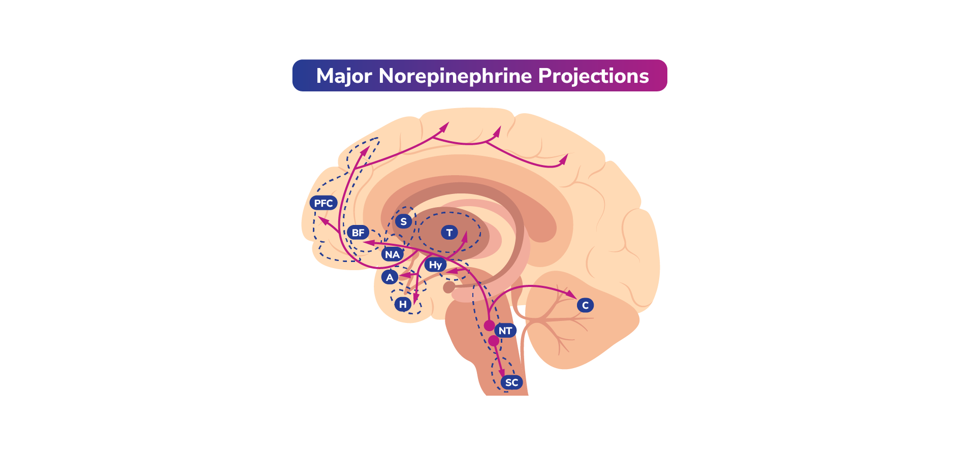
    Norepinephrine, dopamine, and serotonin often work together. Dysfunction of these neurotransmitters is hypothesized to play a role in the symptoms associated with mood disorders. Thus, certain medications utilized for psychiatric conditions such as MDD or bipolar disorder can often impact one or more of these neurotransmitter pathways.1

    The interactions between dopamine and serotonin are of particular importance in helping to regulate mood, cognition, and motor function and will be discussed here. Specifically, activation or inhibition of serotonin receptors can modulate the release of dopamine downstream. 

    5-HT1A Receptors and Dopamine

    It is theorized that the activation of postsynaptic 5-HT1A receptors in the cortex indirectly stimulates dopamine release in the striatum. This is thought to occur through an interplay of reduced glutamate and GABA release, and ultimately a disinhibition of dopamine neurons, leading to dopamine release. Presynaptic 5-HT1A receptors can appear on the dendrites and cell bodies of serotonin neurons within the midbrain raphe and act as autoreceptors. When serotonin is detected at the 5-HT1A receptor, serotonin release is inhibited.1

    5-HT2A Receptors and Dopamine

    The activation of 5-HT2A receptors is believed to indirectly inhibit dopamine release in the striatum. Conversely, blocking 5-HT2A receptors may stimulate dopamine release.1

    Therefore, it is thought that either activating 5-HT1A or blocking 5-HT2A serotonin receptors may lead to the stimulation of dopamine release downstream.1

    5-HT2B Receptors and Dopamine

    Clinical studies have shown that 5-HT2B receptors may be involved in the regulation of basal and “stimulated” dopamine activity, with the ability to regulate dopamine release, dopamine transmission, and dopamine-dependent behaviors.39

    5-HT2C Receptors and Dopamine

    Stimulating 5-HT2C receptors may result in the inhibition of dopamine release, particularly from the mesolimbic pathway. Blocking 5-HT2C receptors may lead to dopamine release in the PFC.

    Mechanisms of Actions and Impact of Medications That May Affect Neurotransmitter Signaling 

    Medications may alter behavior by reducing or enhancing activity at the receptor.

    Agonists (↑): Agonists are compounds that bind to receptors and activate them to the full extent. They can mimic the action of the neurotransmitter and can be used to increase neurotransmitter activity when it is deficient.1

    Antagonists (X): Antagonists bind to receptors but do not activate them. Instead, they block the receptor and prevent it from being activated by agonists or neurotransmitters, thus blocking the agonist-mediated response. Antagonists can be used to inhibit excessive neurotransmitter activity or block unwanted effects of agonists.1

    Partial Agonists (~): Partial agonists can act as functional agonists or functional antagonists depending on the surrounding levels of naturally occurring neurotransmitters. For example, in the absence of a full agonist, a partial agonist can demonstrate agonist activity and activate the receptor. However, the response elicited by a partial agonist will be lower than that of a full agonist. In the presence of a full agonist, however, a partial agonist can act as an antagonist, preventing the receptors from being activated.40

    Partial agonists have a lower intrinsic activity than full agonists. When they bind to receptors, they produce a response that is less than that of a full agonist. The activity of partial agonists at a receptor lies between that of agonists and antagonists.40 Partial agonists can be useful in situations where a full response is not desired.

    Example Actions and Potential Downstream Effects of Drugs Modulating Dopamine and Serotonin Activity  

    Medications may alter behavior by reducing or enhancing activity at the receptor.    Agonists (↑): Agonists are compounds that bind to receptors and activate them to the full extent. They can mimic the action of the neurotransmitter and can be used to increase neurotransmitter activity when it is deficient.1  Antagonists (X): Antagonists bind to receptors but do not activate them. Instead, they block the receptor and prevent it from being activated by agonists or neurotransmitters, thus blocking the agonist-mediated response. Antagonists can be used to inhibit excessive neurotransmitter activity or block unwanted effects of agonists.1  Partial Agonists (~): Partial agonists can act as functional agonists or functional antagonists depending on the surrounding levels of naturally occurring neurotransmitters. For example, in the absence of a full agonist, a partial agonist can demonstrate agonist activity and activate the receptor. However, the response elicited by a partial agonist will be lower than that of a full agonist. In the presence of a full agonist, however, a partial agonist can act as an antagonist, preventing the receptors from being activated.40  Partial agonists have a lower intrinsic activity than full agonists. When they bind to receptors, they produce a response that is less than that of a full agonist. The activity of partial agonists at a receptor lies between that of agonists and antagonists.40 Partial agonists can be useful in situations where a full response is not desired.1   Example Actions and Potential Downstream Effects of Drugs Modulating Dopamine and Serotonin Activity

    Dopamine and Serotonin Receptor Antagonism

    EPS Modulation

    Antagonism of both D2 and 5-HT2A receptors can release the inhibition on dopamine release and lead to an increase in dopamine concentration in the striatum. Increased dopamine release in the striatum due to 5-HT2A antagonism can allow dopamine to compete with the D2 receptor antagonists. This may help reduce the risk of EPS. Conversely, activation of 5-HT1A receptors and the resulting release of dopamine in the striatum is believed to play a role in mitigating the potential EPS that may occur due to D2 antagonism.1

    Prolactin Regulation

    Serotonin and dopamine have opposite roles in regulating prolactin secretion. Dopamine can inhibit prolactin release by stimulating the D2 receptors. Serotonin promotes prolactin release via activation of the 5-HT2A receptors. If the D2 receptors are antagonized, dopamine binding may be disrupted, and prolactin levels may increase. However, if 5-HT2A receptors are blocked in addition to D2 receptors, this may increase dopamine levels, inhibit prolactin release, and potentially help reduce the hyperprolactinemia caused by the D2 receptor blockade alone.

    General Clinical Safety and Monitoring Considerations for Psychopharmacologic Agents

    It is important to note that there are patient-dependent factors that may affect response to psychopharmacological treatment.  

    Monitoring Considerations for Psychopharmacologic Agents

    Drug-drug interaction (DDI)

    For patients that may be taking more than 1 psychotropic medication, adverse events due to DDIs, if they do occur, can often take place within 1 to 2 weeks after a change in existing medication or with the use of an additional medication. Therefore, it is important to include DDIs as a part of differential diagnosis and monitoring.50

    Food-drug interactions

    Pharmacodynamic: Consuming tyramine-rich foods, such as fermented items, while taking a monoamine oxidase inhibitor (MAOI) can lead to a hypertensive crisis. MAOIs prevent the breakdown of both endogenous and dietary amines, including tyramine, a precursor to catecholamines, which are natural vasoconstrictors. By reducing tyramine breakdown and increasing catecholamine production, MAOIs can trigger a dangerous rise in blood pressure.51

    Pharmacokinetic/Absorption: Pairing lipid-soluble drugs with a high-fat meal can lead to increased absorption, resulting in a higher concentration of the drug in the bloodstream. This can increase the potential risk of side effects and toxicity.51

    Grapefruit juice is one of the most extensively studied dietary substances known to inhibit CYP3A and CYP1A2, increasing the half-life of the drug in the blood, which may increase toxicity risk. Additionally, grapefruit juice can act on drug transporter proteins, inhibiting transporter proteins of organic cations and anions.51

    *Tip: Use online drug interaction resources to determine the potential of a DDI when prescribing medications, especially psychotropic medications.50

    Practitioner awareness of potential drug interactions is important. Being able to recognize medications that are commonly associated with narrow therapeutic windows, nonlinear pharmacokinetics, and long half-lives may be beneficial.50

    Patient Monitoring

    Monitor elderly patients, patients with renal or hepatic impairment, and those with symptoms of confusion or sedation closely, as they could be more sensitive to adverse effects or DDIs than other patients.50

    Patients should also closely monitor themselves for adverse effects of drugs with a wide therapeutic window if a specific clinical concentration is not being targeted.52

    Many medication errors can be prevented. A medication history provides information on what a patient has taken in the past and/or is currently taking as well as their response to or side effects of the medication(s). It also gives the provider an opportunity to educate patients about their medications and ensure appropriate use. A proper medication history includes current prescribed drugs, formulations, doses, routes of administration, frequencies, duration of treatment, natural remedies, vitamins, supplements, past medications, previous hypersensitivity and/or adverse reactions (including nature, time course), as well as adherence to treatment.53 

    References

    1. Stahl SM. Stahl’s Essential Psychopharmacology. 4th ed. Cambridge University Press; 2013:89-96.

    2. Teleanu RI, Niculescu AG, Roza E, Vladâcenco O, Grumezescu AM, Teleanu DM. Neurotransmitters-key factors in neurological and neurodegenerative disorders of the central nervous system. Int J Mol Sci. 2022;23(11):5954. doi:10.3390/ijms23115954
    3. Herculano-Houzel S. The human brain in numbers: a linearly scaled-up primate brain. Front Hum Neurosci. 2009;3:31. doi:10.3389/neuro.09.031.2009
    4. Leonard BE. Fundamentals of Psychopharmacology. 3rd ed. John Wiley & Sons, Ltd; 2003.
    5. Hyman SE. Neurotransmitters. Curr Biol. 2005;15(5):R154-R158.
    6. Johnson RL, Koerner JF. Excitatory amino acid neurotransmission. J Med Chem. 1988;31(11):2057-2066.
    7. Tiedje KE, Stevens K, Barnes S, Weaver DF. Beta-alanine as a small molecule neurotransmitter. Neurochem Int. 2010;57(3):177-188. doi:10.1016/j.neuint.2010.06.001
    8. Citri A, Malenka RC. Synaptic plasticity: multiple forms, functions, and mechanisms. Neuropsychopharmacology. 2008;33(1):18-41. doi:10.1038/sj.npp.1301559
    9. Ochoa-de la Paz LD, Gulias-Cañizo R, D´Abril Ruíz-Leyja E, Sánchez-Castillo H, Parodí J. The role of GABA neurotransmitter in the human central nervous system, physiology, and pathophysiology. Revista Mexicana de Neurociencia. 2021;22(2):5355. doi:10.24875/RMN.20000050
    10. Baik JH. Stress and the dopaminergic reward system. Exp Mol Med. 2020;52(12):1879-1890. doi:10.1038/s12276-020-00532-4
    1. Beaulieu JM, Gainetdinov RR. The physiology, signaling, and pharmacology of dopamine receptors. Pharmacol Rev. 2011;63(1):182-217. doi:10.1124/pr.110.002642
    2. Kaur S, Singh S, Jaiswal G, et al. Pharmacology of dopamine and its receptors. In: Kumar P, Deb PK, eds. Frontiers in Psychopharmacology. Springer, Singapore; 2020:143–182. doi:10.1007/978-981-15-3556-7_5
    3. Nichols CD, Amara SG, Sibley R. 5-hydroxytryptamine (serotonin) and dopamine. In: Brunton LL, Knollmann BC, eds. Goodman & Gilman's: The Pharmacological Basis of Therapeutics. 14th ed. McGraw-Hill Education; 2023.
    4. Stahl SM. Dazzled by the dominions of dopamine: clinical roles of D3, D2, and D1 receptors. CNS Spectr. 2017;22(4):305-311. doi:10.1017/S1092852917000426
    5. Adams M, Holland N, Urban C. Pharmacology for Nurses: A Pathophysiologic Approach. 4th ed. Pearson Education, Inc; 2014.
    6. Sokoloff P, Le Foll B. The dopamine D3 receptor, a quarter century later. Eur J Neurosci. 2017;45(1):2-19. doi:10.1111/ejn.13390
    7. Loiseau F, Millan MJ. Blockade of dopamine D(3) receptors in frontal cortex, but not in sub-cortical structures, enhances social recognition in rats: similar actions of D(1) receptor agonists, but not of D(2) antagonists. Eur Neuropsychopharmacol. 2009;19(1):23-33. doi:10.1016/j.euroneuro.2008.07.012
    8. Lin SH, Lee LT, Yang YK. Serotonin and mental disorders: a concise review on molecular neuroimaging evidence. Clin Psychopharmacol Neurosci. 2014;12(3):196-202. doi:10.9758/cpn.2014.12.3.196
    9. Leonard BE. Fundamentals of Psychopharmacology. 3rd ed. John Wiley & Sons, Inc; 2003.
    10. Berger M, Gray JA, Roth BL. The expanded biology of serotonin. Annu Rev Med. 2009;60:355-366. doi:10.1146/annurev.med.60.042307.110802
    11. Mitroshina EV, Marasanova EA, Vedunova MV. Functional dimerization of serotonin receptors: role in health and depressive disorders. Int J Mol Sci. 2023;24(22):16416. doi:10.3390/ijms242216416
    12. Lin J, Liu W, Guan J, et al. Latest updates on the serotonergic system in depression and anxiety. Front Synaptic Neurosci. 2023;15:1124112. doi:10.3389/fnsyn.2023.1124112
    13. Carhart-Harris RL, Nutt DJ. Serotonin and brain function: a tale of two receptors. J Psychopharmacol. 2017;31(9):1091-1120. doi:10.1177/0269881117725915
    14. Wang L, Zhou C, Zhu D, et al. Serotonin-1A receptor alterations in depression: a meta-analysis of molecular imaging studies. BMC Psychiatry. 2016;16(1):319. doi:10.1186/s12888-016-1025-0
    15. Zhang G, Stackman RW Jr. The role of serotonin 5-HT2A receptors in memory and cognition. Front Pharmacol. 2015;6:225. doi:10.3389/fphar.2015.00225
    16. Ebdrup BH, Rasmussen H, Arnt J, Glenthøj B. Serotonin 2A receptor antagonists for treatment of schizophrenia. Expert Opin Investig Drugs. 2011;20(9):1211-1223.
    17. Messa C, Colombo C, Moresco RM, et al. 5-HT2A receptor binding is reduced in drug-naive and unchanged in SSRI-responder depressed patients compared to healthy controls: a PET study. Psychopharmacology. 2003;167(1):72-78. doi:10.1007/s00213-002-1379-5
    18. Pitychoutis PM, Belmer A, Moutkine I, Adrien J, Maroteaux L. Mice lacking the serotonin Htr2B receptor gene present an antipsychotic-sensitive schizophrenic-like phenotype. Neuropsychopharmacology. 2015;40(12):2764-2773. doi:10.1038/npp.2015.126
    19. Popova NK, Tsybko AS, Naumenko VS. The implication of 5-HT receptor family members in aggression, depression and suicide: similarity and difference. Int J Mol Sci. 2022;23(15):8814. doi:10.3390/ijms23158814
    20. Fletcher PJ, Higgins GA. Serotonin and reward-related behavior: focus on 5-HT2C receptors. In: Di Giovanni G, Esposito E, Di Matteo V, eds. 5-HT2C Receptors in the Pathophysiology of CNS Disease. Springer Nature; 2011.
    21. Demchenko I, Tassone VK, Kennedy SH, Dunlop K, Bhat V. Intrinsic connectivity networks of glutamate-mediated antidepressant response: a neuroimaging review. Front Psychiatry. 2022;13:864902. doi:10.3389/fpsyt.2022.864902
    22. Shepherd GM. Corticostriatal connectivity and its role in disease. Nat Rev Neurosci. 2013;14(4):278-291. doi:10.1038/nrn3469
    23. Gómez-Ocádiz R, Silberberg G. Corticostriatal pathways for bilateral sensorimotor functions. Curr Opin Neurobiol. 2023;83:102781. doi:10.1016/j.conb.2023.102781
    24. Calabresi P, Picconi B, Tozzi A, Ghiglieri V, Di Filippo M. Direct and indirect pathways of basal ganglia: a critical reappraisal. Nat Neurosci. 2014;17(8):1022-1030. doi:10.1038/nn.3743
    25. Milardi D, Quartarone A, Bramanti A, et al. The cortico-basal ganglia-cerebellar network: past, present and future perspectives. Front Syst Neurosci. 2019;13:61. doi:10.3389/fnsys.2019.00061
    26. Waldvogel HJ, Baer K, Faull RLM. Distribution of GABAA receptor subunits in the human brain. In: Monti J, Pandi-Perumal S, Möhler H, eds. GABA and Sleep. Springer, Basel; 2010:73-93. doi:10.1007/978-3-0346-0226-6_3
    27. Nuss P. Anxiety disorders and GABA neurotransmission: a disturbance of modulation. Neuropsychiatr Dis Treat. 2015;2015(11):165-175. doi:10.2147/NDT.S58841
    28. Naffaa MM, Hung S, Chebib M, Johnston GAR, Hanrahan JR. GABA-ρ receptors: distinctive functions and molecular pharmacology. Br J Pharmacol. 2017;174(13):1881-1894. doi:10.1111/bph.13768
    29. Devroye C, Cathala A, Piazza PV, Spampinato U. The central serotonin 2B receptor as a new pharmacological target for the treatment of dopamine-related neuropsychiatric disorders: rationale and current status of research. Pharmacol Ther. 2018;181:143-155. doi:10.1016/j.pharmthera.2017.07.014
    30. Lieberman JA. Dopamine partial agonists: a new class of antipsychotic. CNS Drugs. 2004;18(4):251-267. doi:10.2165/00023210-200418040-00005
    31. Choi J, Horner KA. Dopamine agonists. In: StatPearls [Internet]. StatPearls Publishing; 2024.
    32. Chokhawala K, Stevens L. Antipsychotic medications. In: StatPearls [Internet]. StatPearls Publishing; 2024. 
    33. Mohr P, Masopust J, Kopeček M. Dopamine receptor partial agonists: do they differ in their clinical efficacy? Front Psychiatry. 2022;12:781946. doi:10.3389/fpsyt.2021.781946
    34. Casey AB, Cui M, Booth RG, Canal CE. “Selective” serotonin 5-HT2A receptor antagonists. Biochem Pharmacol. 2022;200:115028. doi:10.1016/j.bcp.2022.115028
    35. Mukherjee S, Skrede S, Milbank E, Andriantsitohaina R, López M, Fernø J. Understanding the effects of antipsychotics on appetite control. Front Nutr. 2022;8:815456. doi:10.3389/fnut.2021.815456
    36. Garcia-Garcia AL, Newman-Tancredi A, Leonardo ED. Psychopharmacology (Berl). 2014;231(4):623–636. doi:10.1007/s00213-013-3389-x
    37. Kaufman J, DeLorenzo C, Choudhury S, Parsey RV. The 5-HT1A receptor in major depressive disorder. Eur Neuropsychopharmacol. 2016;26(3):397-410. doi:10.1016/j.euroneuro.2015.12.039
    38. Keks N, Hope J, Schwartz D, McLennan H, Copolov D, Meadows G. Comparative tolerability of dopamine D2/3 receptor partial agonists for schizophrenia. CNS Drugs. 2020;34(5):473-507. doi:10.1007/s40263-020-00718-4
    39. Citrome L, Stensbøl TB, Maeda K. The preclinical profile of brexpiprazole: what is its clinical relevance for the treatment of psychiatric disorders? Expert Rev Neurother. 2015;15(10):1219-1229. doi:10.1586/14737175.2015.1086269
    40. English BA, Dortch M, Ereshefsky L, Jhee S. Clinically significant psychotropic drug-drug interactions in the primary care setting. Curr Psychiatry Rep. 2012;14(4):376-390. doi:10.1007/s11920-012-0284-9
    41. Choi JH, Ko CM. Food and drug interactions. J Lifestyle Med. 2017;7(1):1-9. doi:10.15280/jlm.2017.7.1.1
    42. Asher GN, Corbett AH, Hawke RL. Common herbal dietary supplement-drug interactions. Am Fam Physician. 2017;96(2):101-107.
    43. Fitzgerald RJ. Medication errors: the importance of an accurate drug history. Brit J Clinical Pharma. 2009;67(6):671-675. doi:10.1111/j.1365-2125.2009.03424.x  

    This resource is intended for educational purposes only and is intended for US healthcare professionals. Healthcare professionals should use independent medical judgment. All decisions regarding patient care must be handled by a healthcare professional and be made based on the unique needs of each patient.   

    ABBV-US-01856-MC, Version 1.0
    Approved 02/2025
    AbbVie Medical Affairs

    Welcome To NP Psych Navigator

    This website is intended for healthcare professionals inside the United States.
    Please confirm that you are a healthcare professional inside the US.

    No, I am not US Healthcare professional

    You are now leaving NP Psych Navigator

    Links to sites outside of NP Psych Navigator are provided as a resource to the viewer. AbbVie Inc accepts no responsibility for the content of non-AbbVie linked sites.

    Redirect to:

    Please to bookmark content